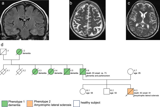
Fig. 1.
Cerebral magnetic resonance imaging (cMRI) in the index patient. cMRI at age 61 years revealed no evidence of atrophy in the frontal lobe, insula or caudate nucleus (a: frontal T2 FLAIR image showing intact insula; b: axial T2-weighted image demonstrating absence of frontal atrophy; c axial T2-weighted image indicating no caudate nucleus atrophy). Pedigree of the index patient. Family history of the index patient (marked by an arrow) is positive for Parkinson`s disease and dementia (d). His father was diagnosed with a “Parkinsonian syndrome” at about age of 60 years, developed symptoms of a dementia with 73 years comprising language decline and changes in behaviour, and died at age 83 years. Two paternal aunts and one paternal uncle out of in total six siblings as well as their mother were also diagnosed with a late-onset neurodegenerative dementia syndrome. None of the relatives were suffering from ALS

