Fig. 2.
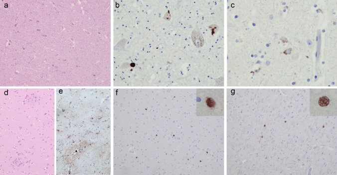
Neuropathology. Typical ALS pathology with (a) loss of motor neurons in the anterior horn of the spinal cord (H&E) and TDP-43-immunoreactive neuronal cytoplasmic inclusions in the spinal cord (b) and precentral gyrus (c). (d) The caudate nucleus revealed no signs of neurodegeneration by H&E. However, mild astrogliosis was detected by GFAP immunohistochemistry (e). PolyQ-immunoreactive neuronal intranuclear inclusions were present in the striatum (f) and frontal cortex (g)

