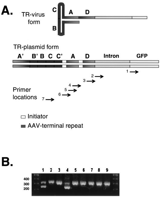
FIG. 2.
(A) Diagram of the TR in genome form as packaged in the virion (top) and as cloned into a plasmid (bottom). The TR forms a T-shaped structure containing one continuous strand in the virion. In the plasmid the TR is double stranded and contains the A, B, and C regions and their complements, A′, B′, and C′. The location within the terminal repeat, intron, or GFP gene of 5′ primers 1 to 7 are also shown. Primer 1 spans −13 to +6 of the GFP gene, primer 2 spans −3 to +15 of the SV40 intron (sequences 5′ of the GFP gene and the intron are polylinker regions), primer 3 spans nt 139 to 145 of the TR plus 15 nt of the polylinker, primer 4 spans nt 110 to 127 of the TR, primer 5 spans nt 104 to 122, primer 6 spans nt 88 to 105, and primer 7 spans nt 60 to 77. (B) RT-PCR products were amplified using primer 2 from RNA isolated from transfected 293 cells and analyzed by agarose gel electrophoresis. Lanes: 1 to 3, TR-pneo (Fig. 1)-transfected cells; 4 to 6, TR/CMV/GFP-transfected cells; 7 to 9, mock-transfected cells that were spiked with TR-pneo DNA at cell harvest; 1, 4, and 7, untreated mRNA; 2, 5, and 8, RNA samples treated with S1 nuclease prior to RT-PCR; 3, 6, and 9, RNA samples treated with NaOH to degrade RNA prior to RT-PCR. The 3′ primer paired with primer 2 was located in the GFP gene: GFP-RT2 (nt 145 to 126 of the EGFP gene). Either GFP-RT2 or GFP-RT1 (nt 19 to 3 of the EGFP gene) was paired with the 5′ primers from panel A, as delineated in Table 1.

