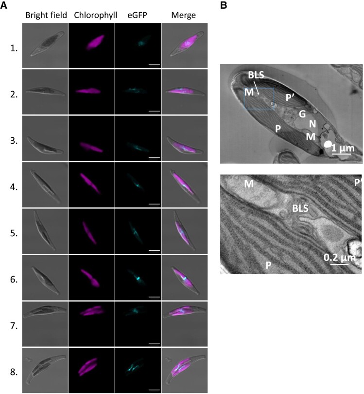
Figure 3.
Subcellular localization of the blob-like structure, associated to MGDβ–eGFP fluorescence. Observation of blob-like structure localization by confocal microscopy (A) and transmission electron microscopy (B). P. tricornutum transgenic lines overexpressing MGDβ–eGFP were used to monitor the appearance of blob-like structures in the cell (A). Cells 1 and 2 show a scattered eGFP signal, Cells 3 to 6 show a blob-like structure at the central constriction of dividing plastids, Cell 7 shows a blob-like structure extending from 1 plastid to the other inside a dividing cell, and Cell 8 shows 2 blob-like structures facing each other on distinct plastids during cytokinesis. Cell 6 was also shown in Fig. 2. Observation of a WT P. tricornutum cell containing 2 plastids following plastid division inside the cell (B). Cleavage furrows are visible at both end of the cell. The blob-like structure (outlined in a rectangle) was observed connecting the 2 plastids at 2 magnifications. BLS, blob-like structure (or blob); G, Golgi apparatus; M, mitochondrion; MGD, monogalactosyldiacylglycerol synthase; N, nucleus; P and P′, plastids.

