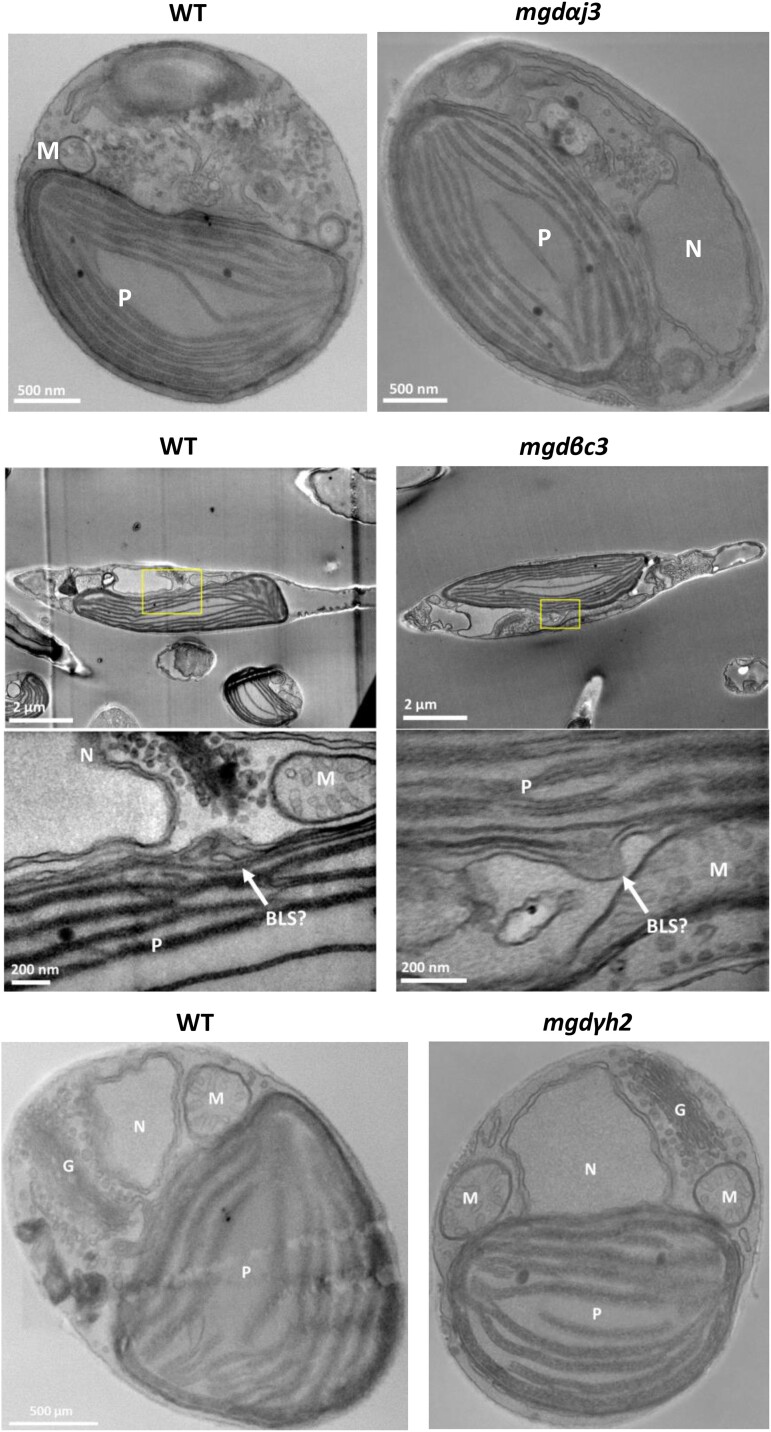
Figure 4.
Phaeodactylum tricornutum cell ultrastructure in MGD KO lines determined by STEM. A WT and KO mgdαj3 and mgdγh2 lines were cultured in parallel. In a separate experiment, a WT and a mgdβc3 mutant were cultured in parallel. Cell ultrastructure is shown in each mutant with corresponding WT control on the left. No impact of MGDα, MGDβ, or MGDγ KO could be observed at the level of membrane compartments, including plastids. BLS outlined in rectangles was observed at 2 magnifications. BLS, blob-like structure; G, Golgi; KO, KO; M, mitochondria; MGD, monogalactosyldiacylglycerol synthase; N, nucleus; P, plastid; WT, wild type.

