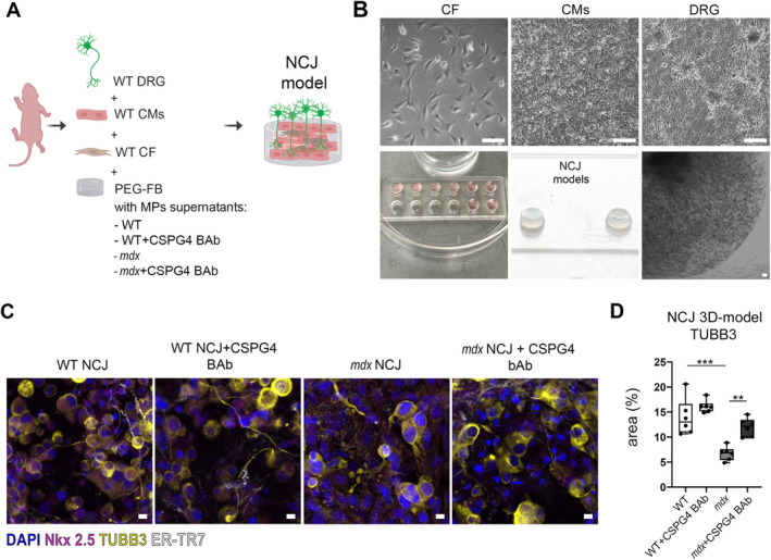
Figure 3.

MPs release CSPG4 in media and inhibit axon growth of dorsal root ganglia. (A) Rendering of 3D NCJ generation. The picture was created with Biorender.com and illustrates the composition of the 3D NCJ model: WT dorsal root ganglia (DRG), WT cardiomyocytes (CMs), and WT CFs were encapsulated in PEG‐FB hydrogel conditioned with the supernatants derived from MP cultures of WT, WT + CSPG4 blocking antibody (BAb), mdx, and mdx + CSPG4 (BAb). (B) Brightfield images of CFs, CMs, and DRG after 1 day of culture (upper panel) and 3D constructs of NCJ models after 7 days (lower panel). Scale bars, 100 μm. (C) Confocal images of 3D NCJ models: WT, WT + CSPG4 BAb, mdx, mdx + CSPG4 BAb. DRG are labeled with tubulin beta 3 (TUBB3, in yellow), CMs with cardiac troponin (cTNNT, in magenta), and CFs with fibroblast marker (ER‐TR7, in white). (D) The graph indicates the area (%) occupied by neuron endings labeled by TUBB3 inside the 3D NCJ models. N = 3 biological replicates, N = 2 sections/constructs. Scale bars, 10 μm. **p < 0.01, ***p < 0.001 were determined using one‐way ANOVA.
