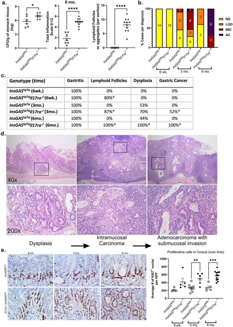
Figure 2.

IL-17RA deficiency leads to increased cellular proliferation and development of gastric cancer in chronically infected mice. A. At the 6 mo. post-infection time point, colonization levels, total inflammation scores and number of lymphoid follicles per section are quantified. B. Frequency of adverse pathological findings in gastric tissue defined by pathologist at 3 defined timepoints post-infection of PMSS1 H. pylori. the numbers of cases per diagnosis are represented within each bar on the graph; diagnoses are defined as: no dysplasia (ND, gastritis only), low grade dysplasia (LGD), intramucosal carcinoma (IMC), and adenocarcinoma with invasion to the submucosa (AC). C. The table also demonstrates frequency of pathological outcomes including lymphoid follicles, dysplasia and gastric cancer. *p < 0.05, Fisher’s exact test was used to test significance of these pathological findings comparing genotypes at each time point. D. Representative H&E-stained gastric sections were imaged and are displayed at 40× (top) and 200× (bottom). Pathological outcomes of dysplasia, intramucosal carcinoma, and invasive adenocarcinoma occur in InsGAStg/tgIL-17ra-/- at 6-month post H. pylori infection and are represented from left to right. E. Ki67 staining and quantification within gastric tissues of H. pylori infected mice at 3 defined time points. Five high power fields (HPF) were counted per case. The average number of counts per case was graphed by genotype and time point (the average from each case is represented by the data point, mean is indicated, error bars represent ± SEM).
