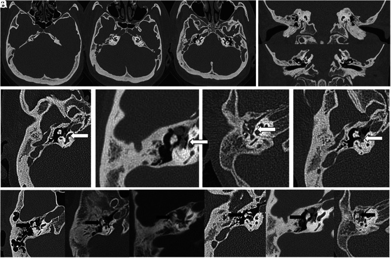
Figure 1.
Axial (A) view of a typical temporal bone computed tomography scan of an achondroplastic patient demonstrating prominent emissary veins, poor mastoid pneumatization, and carotid canal shortening. Coronal (B) view of a typical temporal bone computed tomography scan of an achondroplastic patient displaying upward tilting of the external and internal auditory canals, downward-facing oval window, and horizontal placement of scutum. (C) Aberrant facial nerve course (white arrow) observed in 4 temporal bone computed tomography scans. The facial nerve overhangs the oval window and eclipses the incudostapedial joint and stapes suprastructure in the axial view. (D) Six temporal bone computed tomography scans showing a hypoplastic and broad-based incus body (black arrow), presumably caused by dysfunction in endochondral ossification.

 Content of this journal is licensed under a