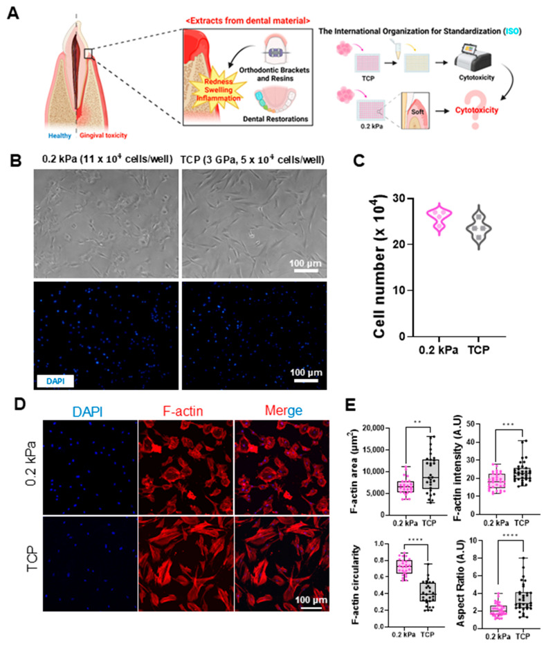
Figure 2.
Impact of dental material extracts on gingival tissue and cellular responses to substrates with different stiffnesses. (A) Schematic representation of the potential cytotoxic effects of dental material extracts on gingival tissue. While current ISO standards utilize TCP (stiff) for testing, dental materials in actual gingival environments interact with soft tissues. This image illustrates the hypothesis that the cytotoxicity of these extracts may differ in soft gingival conditions compared to standard stiff substrates. (B) Representative bright-field (top) and DAPI-stained fluorescent images (bottom) of human gingival fibroblasts (HGFs) cultured on 0.2 kPa and TCP (3 GPa) substrates and seeded at 11 × 104 cells/well and 5 × 10⁴ cells/well, respectively. Scale bar = 100 μm. (C) Quantitative comparison of HGF counts on 0.2 kPa and TCP after 24 h incubation, showing minimal differences (n = 4). (D) Immunofluorescent staining of DAPI (blue) and F-actin (red) in HGFs on each substrate. Scale bar = 100 μm. (E) Quantitative analysis of F-actin area, intensity, circularity, and aspect ratio. Cells on 0.2 kPa showed smaller areas, decreased intensity, and a more rounded morphology compared to TCP (n = 30). Data are mean ± SD from three independent experiments. Statistical significance was determined by Student’s t-test (** p < 0.01, *** p < 0.001, **** p < 0.0001).

