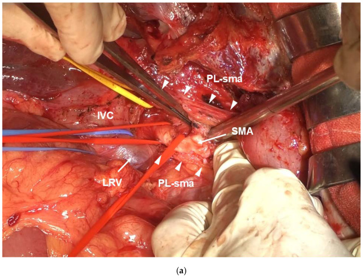
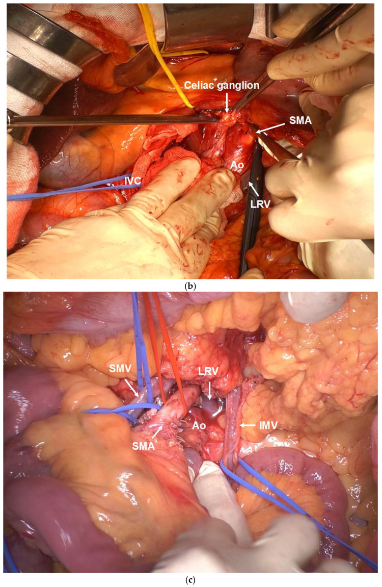
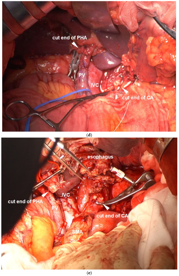
Figure 3.
(a) In the visual field of Kocher mobilization, the SMA plexus (Δ) is opened dorsally in a double-door manner across approximately 7 cm from the SMA root. (b) The right celiac plexus and right celiac ganglion are separated to reach the anterior aspect of the Ao. (c) The mesenteric approach allows observation of the entire SMA length from the ventral side. (d) Photograph of the completed resection by DP-CAR. The CA stump is clamped with vascular clamping forceps, leaving a suture margin required for anastomosis. (e) Photograph of the completed resection using TP-CAR+TG. Wide field of view because of en bloc dissection of the left upper abdominal organs.


