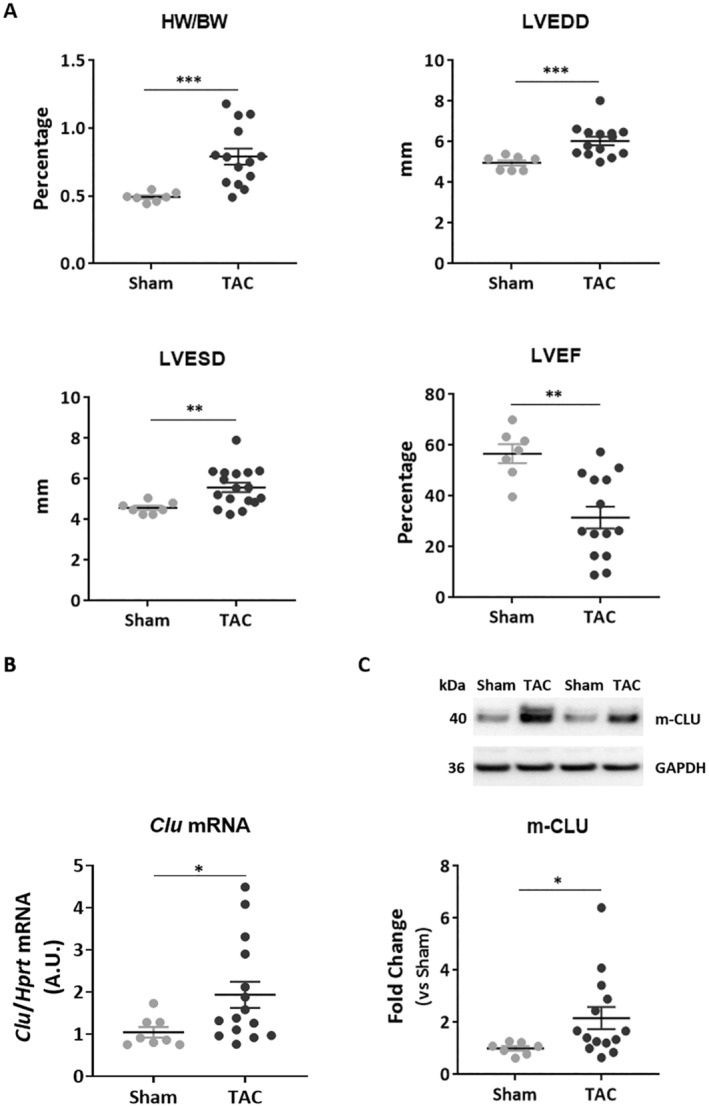
FIGURE 1.

CLU expression is induced in the heart after transverse aortic constriction (TAC). (A) Animal characteristics after 6–8 weeks of TAC were quantified for heart weight/body weight (HW/BW) ratio, left ventricle end diastolic diameter (LVEDD), left ventricle end systolic diameter (LVESD), and ejection fraction (EF) in sham (n = 7) and TAC mice (n = 14). (B) Quantification of Clu mRNA level by qPCR in the left ventricle of sham and TAC mice. Hprt was used for normalisation. (C) Representative image and quantification of CLU mature protein form (m‐CLU) by western blot in the same samples. GAPDH was used for normalisation. Statistical significance was determined by the Wilcoxon‐Mann–Whitney test. *p < 0.05, **p < 0.01, and ***p < 0.001.
