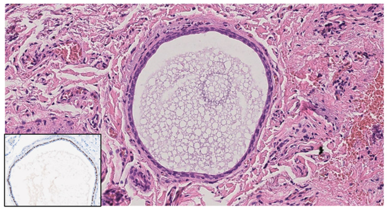
Figure 3.
Histological image. Dermal cyst is composed of an inner layer of cuboidal epithelium and an outer myoepithelial cell layer; p63 (insert) highlights myoepithelial cells. Hematoxylin and Eosin; original magnification (OM) ×20; immunohistochemistry, chromogen diaminobenzidine (insert); original magnification (OM): p63, ×20.

