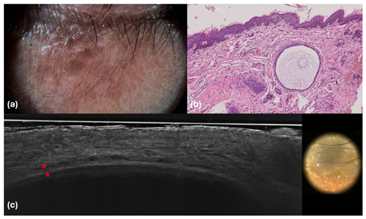
Figure 4.
Dermoscopic (a), histological (b) and LC-OCT (c) images. (a) Purple-bluish color; (b) dermal cystic lesion; (c) hyporeflective cupuliform area well defined by a bright and thick upper contour with two bright layers of cells (red asterisks). Hematoxylin and Eosin; original magnification ×20.

