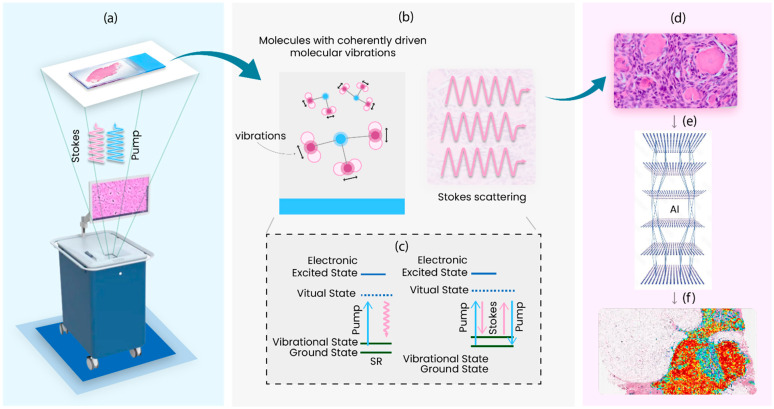
Figure 1.
Overview of SRH workflow. (a) The tumor specimen obtained intra operatively is loaded onto slides and SRH imaging is performed. Stokes and pump lasers illuminate the sample and (b) induced molecular vibrations within the sample. The laser excitation causes energy transitions as shown in (c). The molecular perturbations produce coherent Raman scattered photons that will be collected and pseudo-colored to generate stimulated Raman histology images, as shown in (d). In (e), the resultant images are processed using advanced AI modalities to identify regions of different pathologic features which are heat mapped, as in (f), for easy processing by pathologists.

