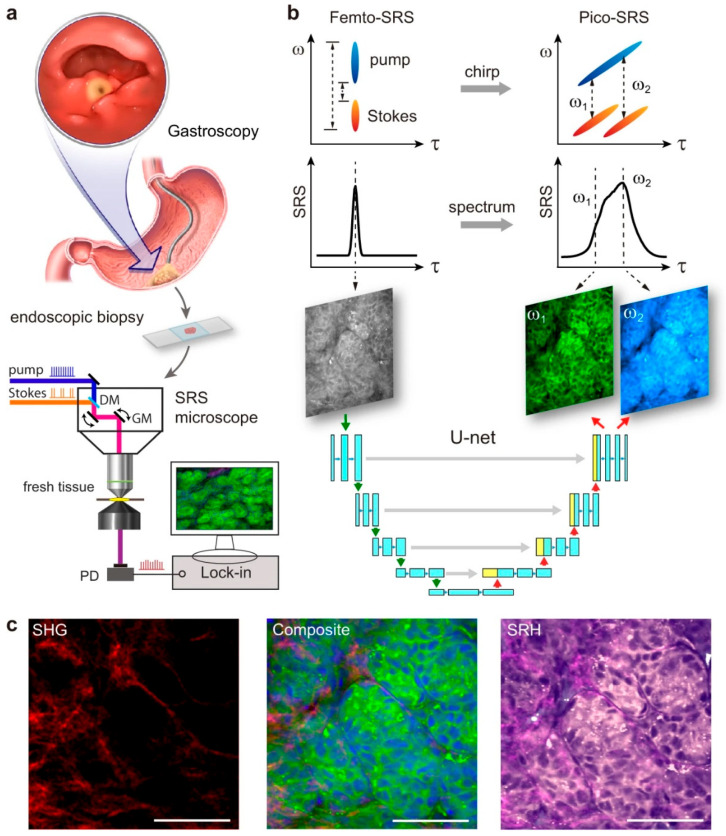
Figure 2.
(a) The representation portrays the process of gastroscopy and the collection of fresh biopsies for direct SRS imaging. (b) It features the properties of Femto-SRS and Pico-SRS, including pulse chirping, spectral resolution and the conversion of a single Femto-SRS image into a pair of Pico-SRS images using deep U-Net. (c) Multi-chemical imaging of gastric tissue including lipid, protein and collagen fibers visualized through converted Femto-SRS and SHG channels, color-coded to SRH. Scale bars: 50 µm. (adopted from Liu et al. [85]).

