Figure 6.
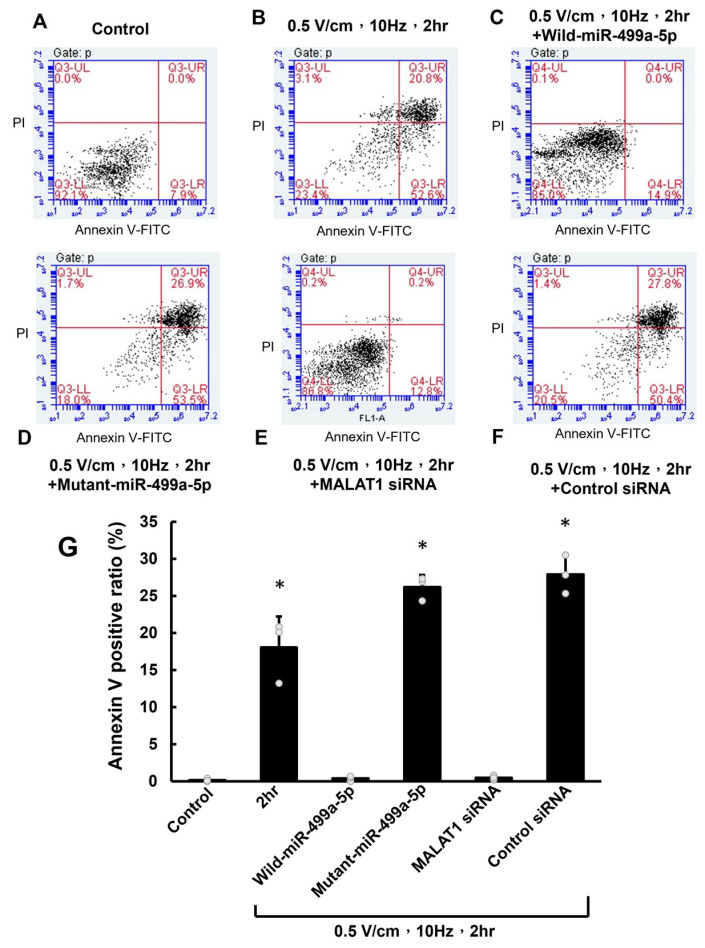
miR-499a-5p and MALAT1 mediate RES-induced apoptosis in HCF-aa. (A,B) Representative flow cytometry plots of propidium iodide–annexin V double-staining in control and RES-treated (0.5 V/cm, 10 Hz, 2 h) HCF-aa. (C) Flow cytometry plot showing the effect of miR-499a-5p overexpression on RES-induced apoptosis. (D) Flow cytometry plot demonstrating the impact of mutant miR-499a-5p on RES-induced apoptosis. (E) Flow cytometry plot showing the effect of MALAT1 siRNA on RES-induced apoptosis. (F) Flow cytometry plot demonstrating the impact of control siRNA on RES-induced apoptosis. (G) Quantitative analysis of apoptotic cells (positive for annexin V and propidium iodide) under all tested conditions. Data represent mean ± SEM from three independent experiments (n = 5 per group). * p < 0.05 vs. control.

