Figure 2.
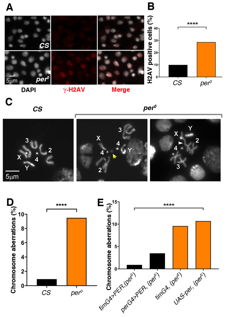
DNA damage and chromosome aberrations in per0 mutants. (A) Anti-γ-H2AV immune labelling (red) in CNS-squash preparations from third-instar male larvae. DNA is stained with DAPI (white). Size bar = 5 μm. ZT = 1. (B) Proportion of cells showing anti-γ-H2AV immune signal in CNS-squash preparations from CS and per0 third-instar male larvae. Cells were considered ‘H2AV positive’ when the relative intensity of the anti-γ-H2AV immune signal in the nucleus [(signal-background)/background] was equal to or more than 1.5. The DAPI signal was used to identify nuclei. Fisher’s exact test, **** p < 0.0001. Total number of cells, n = 1323, 985, respectively. ZT = 1. (C) Mitotic metaphases in CNS-squash preparations from third-instar larvae. Left, CS, showing a normal metaphase. Right, chromosome aberrations in per0. The yellow arrow indicates a chromosome fragment (break). The white arrow points to a fusion. Numbers 2, 3, 4 identify the autosomes. X, Y label the sex chromosomes. Size bar = 5 μm. ZT = 2. (D) Frequency of chromosome aberrations [(abnormal metaphases/total metaphases) × 100] in CS and per0. Fisher’s exact test, **** p < 0.0001. ZT = 2. Total number of metaphases scored, N = 436, 527, respectively. (E) PER overexpression rescues chromosome aberrations in per0. The overexpression of PER using the pan-circadian per-GAL4 (perG4 > PER, per0) and tim-GAL4 (timG4 > PER, per0) drivers drastically reduced the frequency of aberrations in an otherwise per0 background. Chi-square = 49.50, df = 3, **** p < 0.0001. Total number of metaphases scored (from left to right), n = 337, 463, 345, 1021. ZT = 2.

