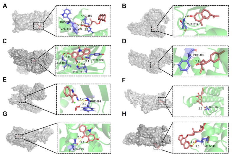
Figure 6.
Molecular docking interactions of various compounds with target proteins. The panels illustrate the binding interactions for different compounds. (A) Wampetin with protein 6m9t; (B) Nootkatone with protein 7jvr; (C) 1,3-Dihydroxy-N-methylacridone with protein 7jvr; (D) (+)-ar-Turmerone with protein 7jvr; (E) 1,2,3,4-Tetrahydro-3-carboxyharmane with protein 7jvr; (F) Myricetin with protein 5iu4; (G) 1,3-Dihydroxy-N-methylacridone with protein 5iu4; (H) 2′,3′-Epoxyindicolactone with protein 5iu4. Each figure presents a 3D macroscopic view of the docking result on the left and a 3D interaction view on the right, with red representing the small molecule compound ligand and blue indicating the amino acid residues.

