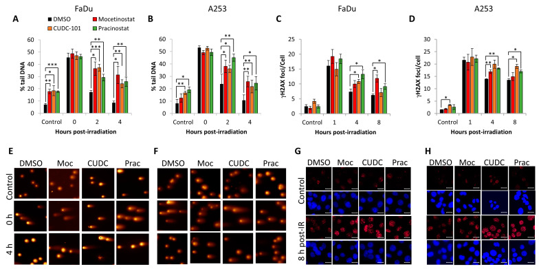
Figure 5.
Mocetinostat, CUDC-101, and pracinostat cause delays in DSB repair in HNSCC cells. (A,C,E,G) FaDu and (B,D,F,H) A253 cells were treated with 1 µM CUDC-101, pracinostat, or DMSO as a control, and either unirradiated (control) or irradiated with 4 Gy X-rays and cells harvested at the various time-points post-irradiation. (A,B) Levels of DSBs were analysed directly using the neutral comet assay, with mean percentage tail DNA ± SE determined from three independent experiments. (C,D) Numbers of γH2AX foci were determined using immunofluorescence microscopy, with mean γH2AX foci/cell ± SE determined from three independent experiments. * p < 0.05, ** p < 0.01, *** p < 0.001 on one-sample t-tests. (E,F) Stained DNA in cells either unirradiated or 4 h post-irradiation following inhibitor treatment through gel electrophoresis. (G,H) γH2AX foci in cells either unirradiated or 8 h post-irradiation following inhibitor treatment. Scale bar is 20 µm.

