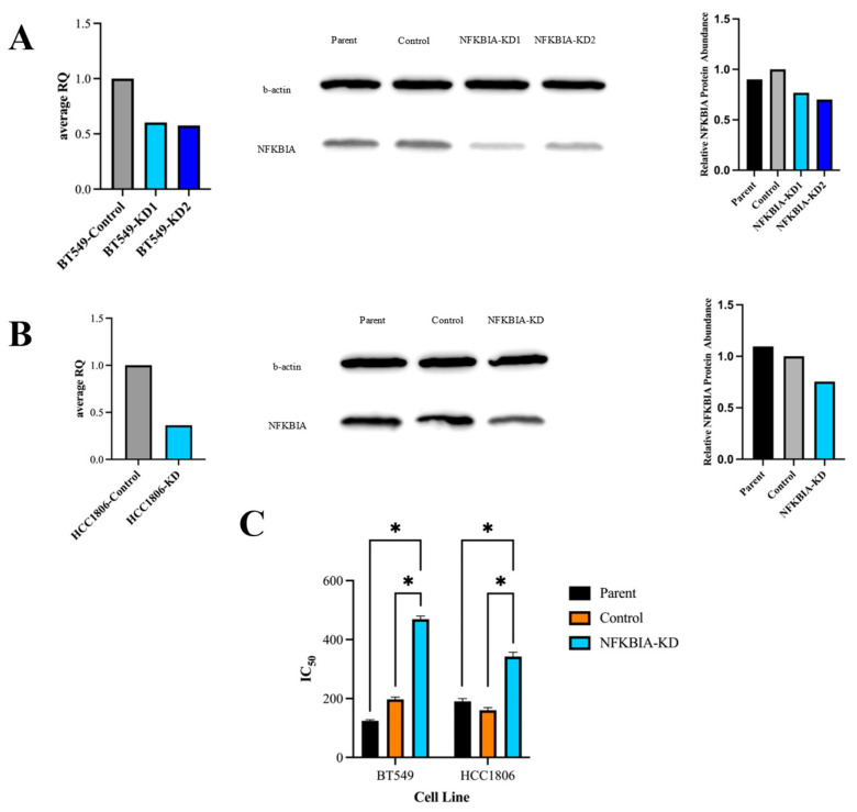
Figure 6.
Knockdown of NFKBIA decreases sensitivity to selinexor. (A) BT-549 NFKBIA gene and protein expression following shRNA knockdown assessed using RT-qPCR (left panel) and Western blot (middle and right panels). Samples of the control scrambled insert as well as NFKBIA knockdown were collected and analyzed at both the gene and protein levels; both confirmed the decreased levels of NFKBIA within the generated knockdown cell line compared to the control scrambled. (B) Similarly to (A), the NFKBIA gene and protein expression following shRNA knockdown in HCC-1806 cells were assessed via RT-qPCR and Western blot. The knockdown-generated version of the cell line showed decreased levels of NFKBIA at both the gene and protein levels. (C) IC50 results for parent, control generated and NFKBIA knockdown-generated cells in both BT-549 and HCC-1806 are shown with standard error bars and factoring in biological replicates. Like Figure 2, 10-point dose–response curves were fit testing the viability of all cell lines after 72 h treatment of between 0 and 3200 nM selinexor. In both cell lines, NFKBIA knockdown displayed statistically significant higher IC50 values compared to either parent or control as indicated by * in (C) (p-value: < 0.0001).

