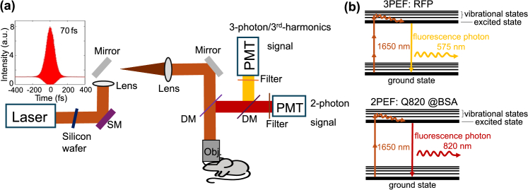
Fig. 1.
Imaging setup and 2-photon and 3-photon excitation schemes at 1650 nm for simultaneous imaging of neurovascular structures. (a) A laser (OPA, Opera-F, Coherent), pumped by an amplifier (Monaco, Coherent), is directed into a multiphoton imaging system (Movable Objective Microscope, Sutter Instrument). Before entering the system, a 3-mm thick silicon wafer is used to compensate for the dispersion of our imaging system. The pulse’s full width at half maximum (FWHM) is ∼70 fs after the objective assuming a sech2(τ) temporal pulse intensity profile. SM: scanning mirrors; DM: dichroic mirrors. Details on the setup are described in the methods section: imaging setup. (b) Simultaneous 1650 nm 3-photon excitation of red fluorescent protein (RFP tdimer2(12)) for labeling neurons and 2-photon excitation of Q820@BSA for labeling vasculature in the brain in vivo.

