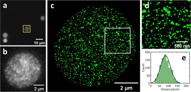
Fig. 6.
Single-color super-resolution imaging results of erythrocytes by SRIF-fluidics. (a) Wide-field fluorescence image of multiple erythrocytes, the cell within the yellow dotted box was selected for the super-resolution imaging. Scale bar: 10 µm. (b) Zoomed-in wide-field image of erythrocytes shown in (a). Scale bar: 2 µm. (c) Super-resolution image of erythrocytes in (b). Scale bar: 2 µm. (d) Enlarged view of image of the area within the white box in (c). Scale bar: 500 nm. (e) Distribution of distances between the nearest neighbors of clusters.

