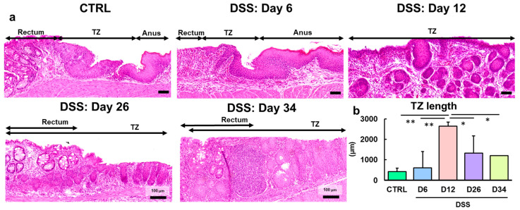
Figure 1.
Morphological characterization of the TZ in the control and DSS-treated groups. (a) Representative images of the TZ in the control mice and mice administered 4% or 5% DSS in drinking water for 6 days (Day 6), followed by withdrawal of DSS for 6 days (Day 12), 20 days (Day 26), and 28 days (Day 34). In the control mice, the TZ with non-keratinizing squamous epithelium was observed between the rectum with crypts and the anus with keratinizing squamous epithelium. In the DSS-treated mice, the non-keratinizing squamous epithelium covered the ulcer lesions in the rectum on Day 6 and proliferated intensely toward the depths on Day 12. On Days 26 and 34, pathological remodeling was shown as mixed with regenerative crypts (light blue triangles) and the hyperplastic TZ (yellow triangles) in the ulcer regions (see Figure S2). Hematoxylin and eosin stain. Bar = 50 (CTRL, Days 6 and 12) or 100 µm (Days 26 and 34). (b) Comparison of TZ lengths in the control and DSS-treated groups at each time point. The mice were treated with 5% DSS for 6 days and euthanized on days 6 and 12. The other mice were treated with 4% DSS for 6 days and euthanized on days 26 and 34. N = 3 (CTRL), 6 (D6), 9 (D12), 3 (D26), and 2 (D34). *, ** Significant differences between each time point or region (p < 0.05 or 0.01, Tukey–Kramer multiple comparison test). CTRL, control; DSS, dextran sodium sulfate; D6, Day 6; D12, Day 12; D26, Day 26; D34, Day 34; TZ, transitional zone.

