Figure 6.
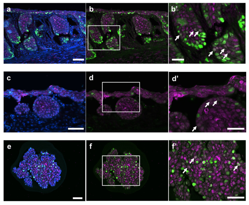
Some SOX2-retaining cells colocalize with SOX9 or LGR5-retaining cells. Representative images of TZ (a–d) and organoids of a non-keratinizing squamous cell (e,f) in the treated mouse administered 4% DSS in drinking water for 6 days, followed by withdrawal of DSS for 6 days (Day 12: (a–d) and 21 days (Day 26; (e,f)). Double immunofluorescence staining for SOX2 (Magenta) and SOX9 (Green) (a,b) or SOX2 (Magenta) and LGR5 (Green) (c,d) in TZ. Double immunofluorescence staining for SOX2 (Magenta) and SOX9 (Green) in the organoid (e,f). Nuclei were counterstained with DAPI (blue). Higher magnification of the region for indicated square (white line) in Figure 6b,d,f), with a white arrow showing colocalization (b’,d’,f’). SOX2 immunoreactivities in mice and organoids derived from TZ were observed in the nucleus as a diffuse pattern. LGR5 and SOX9 were mainly observed in the nucleus for a marginal region at TZ. The nuclei colocalized with SOX2 and SOX9 (SOX2+SOX9low cells) or SOX2 and LGR5 (SOX2+LGR5low cells) were shown as arrows (b’,d’,f’). Bar = 50 µm ((a–f) are the same sections) or 25 µm (b’,d’,f’). DSS, dextran sodium sulfate; LGR5, leucine-rich repeat-containing G-protein-coupled receptor 5; SOX2, sex-determining region on Y-box transcription factor 2; SOX9, sex-determining region on Y-box transcription factor 9; TZ, transitional zone.

