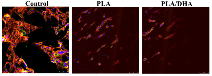
Figure 6.
The immunofluorescence staining of fibroblasts cultured on tissue culture polystyrene (TCPS) (control) and a PLA or a PLA/DHA core–shell fibrous membrane (CSFM). Actin cytoskeleton arrangement (red) and the expression of the focal adhesion protein vinculin (green) on day 3 were examined under a laser confocal microscope after counterstaining the nuclei (blue). Bar = 75 μm.

