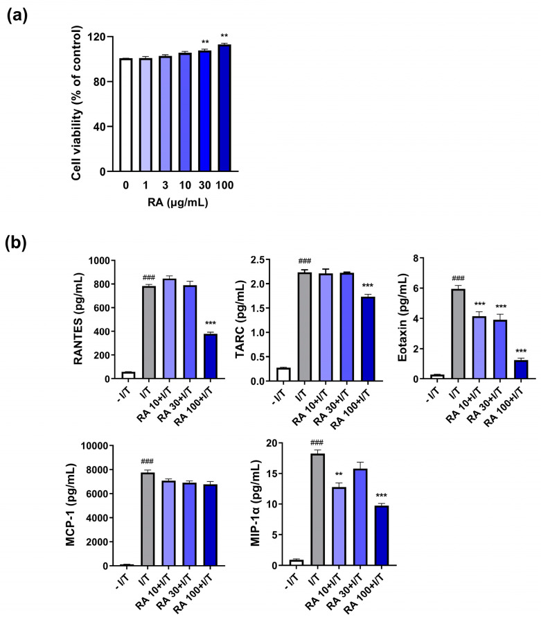
Figure 3.
RA attenuated inflammatory-related chemokine levels in IFN-γ/TNF-α-stimulated HaCaT cells. (a) The cells were treated with RA (1, 3, 10, 30, or 100 µg/mL) for 24 h, and cell viability was measured by an CCK-8 assay. (b) The levels of five inflammatory chemokines (RANTES, TARC, eotaxin, MCP-1, and MIP-1α) in the media were measured via ELISA as described in the Section 4. ### p < 0.001 vs. the vehicle-treated cells without IFN-γ/TNF-α (– I/T) (10 ng/mL); ** p < 0.01 and *** p < 0.001 vs. the vehicle-treated cells with IFN-γ/TNF-α (I/T).

