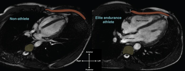
Figure 3.
The athlete's heart within the thorax. Horizontal long axis cardiac MRI of a non-athlete (left) and elite endurance athlete’s heart (right), both 24-year-old males. Compared to the non-athlete, note the significantly greater proportion of the thorax inhabited by the athlete’s heart and the intimate relationship between the right ventricle and the chest wall (anterior, rust) and the left atrium and thoracic vertebrae (posterior, yellow). The right ventricle and left atrium of the athlete’s heart appear to be deformed in shape compared to the non-athlete.

