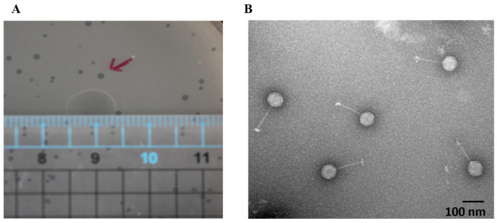
Figure 1.
JSSK01 morphology under transmission electron microscopy. (A) JSSK01 plaque formation in a plaque assay measuring 0.1–0.15 cm; the red arrows represent typical plaques. (B) JSSK01 phage is classified as a siphophage with a 65 ± 4 nm (n = 10) icosahedral head and a 124 ± 6 nm (n = 10) nm long tail, electron micrograph of JSSK01 virions with scale bar of 100 nm, viewed at 150,000 × magnification under negative staining with 2% uranyl acetate.

