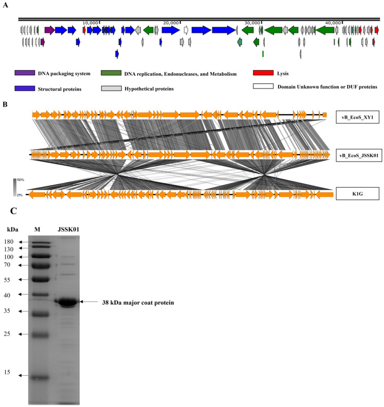
Figure 3.
JSSK01 genome organization, comparison, and structural protein analysis. (A) Each arrow represents an open reading frame (ORF). Based on their encoded protein function, a color was assigned to each ORF. (B) The JSSK01 phage genome was compared via Easyfig with those of two other similar phages, Escherichia siphophages XY1 and K1G. (C) The protein structure of JSSK01 was analyzed via sodium dodecyl sulfate-polyacrylamide gel electrophoresis. The gel was visualized via an iBright imaging system, and the major coat protein was identified at 38 kDa and confirmed via tandem mass spectrometry.

