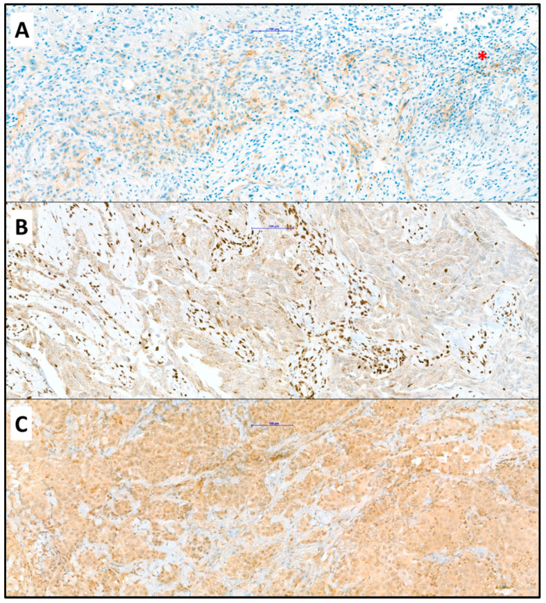
Figure 1.
(A) Positive reaction of mesothelioma cells to antibody against PD-L1 with few positive lymphocytes (asterisk); (B) Loss of nuclear staining in mesothelioma cells with unspecific granular cytoplasmic reaction and clearly positive nuclear staining of lymphocytes (positive internal control); (C) Positive reaction of mesothelioma cells to antibody against ILK; (Objective ×20, immunohistochemistry). The blue bar indicates 100 µm.

