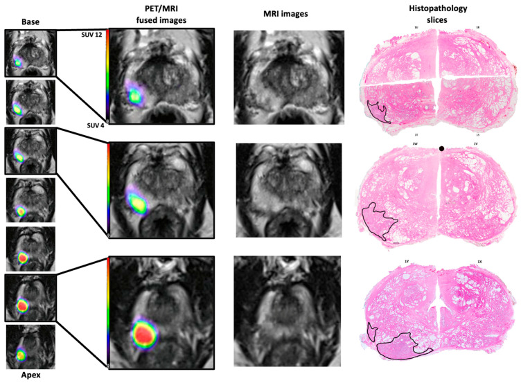
Figure 1.
PET/MRI fused images qualitatively compared to T2WI axial slices of MRI and digitally recreated histopathology slices for a patient from the MRI−/PET+ group. Note the similar localisation of the tumour: right posterior from base to apex. The histology slices are digitally recreated from multiple histology slides and the tumour outlines (black) performed by an experienced uro-pathologist. Note the spacing between the fused images is 3 mm whereas the histopathology slides are approximately 10 mm.

