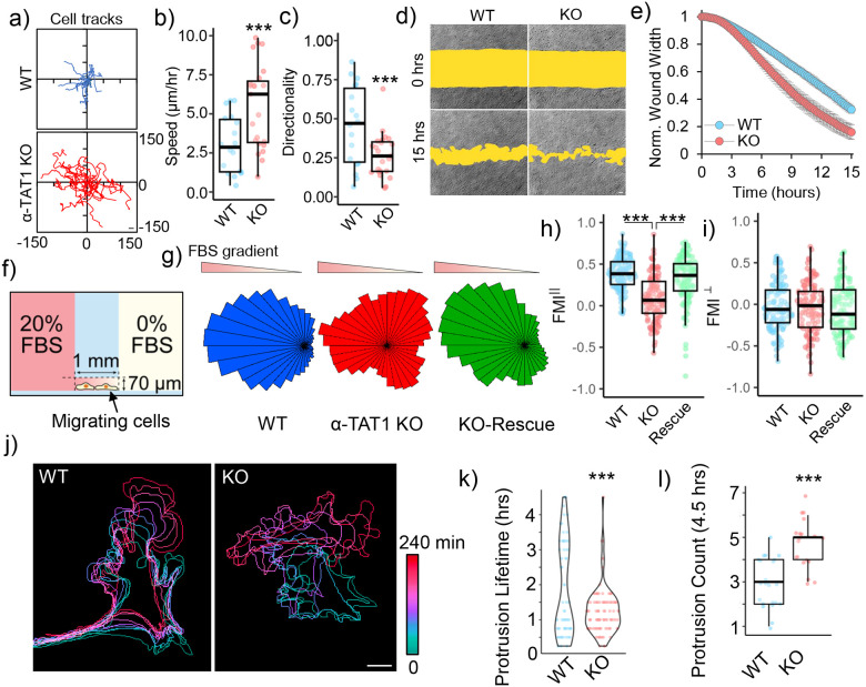
Figure 1. α-TAT1 modulates directional cell migration.
a) Tracks, b) Speed (μm/hr) and c) Directionality of WT and α-TAT1 KO MEFs in a random migration assay, WT: 18, KO: 23 cells, scale bar: 10 μm; d), e) Temporal changes in wound width in a wound healing assay with WT and α-TAT1 KO MEFs, n = 12 wound regions from 3 independent experiments, mean ± 95% C.I.; f) Schematic for chemotaxis assay (adapted from Ibidi); g) Rose plots of WT, α-TAT1 KO or KO-rescue MEFs migrating in a chemotactic gradient, h) Forward migration indices along the chemotactic gradient and i) Forward migration indices perpendicular to the chemotactic gradient for WT, α-TAT1 KO or KO-rescue MEFs, n = 120 cells (40 each from three independent experiments); j) Temporal changes in morphology of WT or α-TAT1 KO MEFs undergoing random migration, scale bar: 10 μm; k) Persistence of protrusions, l) Frequency of new protrusion formations in randomly migrating WT or α-TAT1 KO MEFs, WT: 23 and KO: 19 cells. ***: p<0.001

