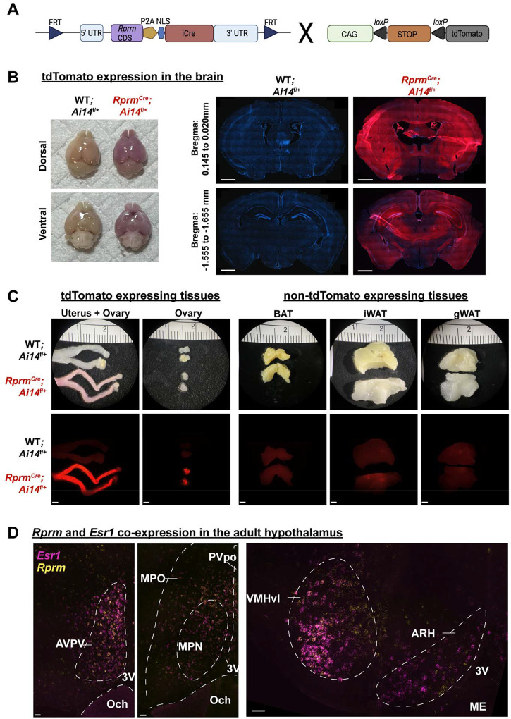
Figure 1: Rprm lineage tracing reveals robust expression in the brain and peripheral tissues.
(A) Schematic representation for the generation of RprmCre;Ai14f/+ mice. Heterozygous RprmCre mice were bred with Ai14f/f mice to produce RprmCre;Ai14 f/+ mice expressing tdTomato in Rprm lineage tissues. Cre-negative littermates (WT;Ai14 f/+ mice) were used to establish baseline tdTomato levels. (B-D) Representative images of tissues harvested from adult (13–15 weeks old) RprmCre;Ai14 f/+ and WT;Ai14 f/+ mice. (B) Left – images highlighting tdTomato expression throughout the whole brain in male mice. Right – 30 μm DAPI-stained coronal brain sections emphasize the expression of tdTomato in the cortex, thalamus, and hypothalamus across representative bregma levels. Scale bars = 1 mm. (C) Left – peripheral tissues exhibiting tdTomato expression include the uterus, & ovary in female mice. Right – adipose tissue depots (BAT, iWAT, and gWAT) do not express tdTomato. Brightfield images displayed on top, fluorescent images below. Scale bars = 200 μm. (D) Single-molecule in situ hybridization reveals Esr1 (magenta) and Rprm (yellow) co-expression in the AVPV, VMHvl, and ARH of an adult female mouse and the MPO of an adult male mouse. Dashed lines indicate regions of interest and landmarks. Scale bars = 50 μm. BAT = brown adipose tissue, iWAT = inguinal white adipose tissue, gWAT = gonadal white adipose tissue, AVPV = anteroventral periventricular nucleus, Och = optic chiasm, 3V = third ventricle, MPO = medial preoptic area, MPN = medial preoptic nucleus, PVpo = Periventricular hypothalamic nucleus, VMHvl = ventrolateral area of ventromedial hypothalamus, ARH = arcuate nucleus, ME = median eminence.

