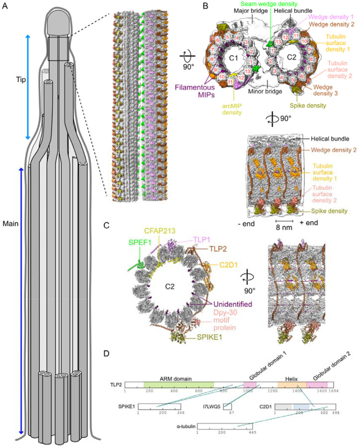
Figure 1: Atomic model of the tip CP.
(A) Cartoon of cilia in Tetrahymena indicating the main and tip regions of the axoneme. The inset shows the cryo-EM map of the distinct structure of the tip CP. (B) Cryo-EM map of the tip CP. Tubulin is shown in gray, and the densities corresponding to the proteins identified in this study are shown in color. The low-resolution map of the tip CP (consensus map) is shown as an envelope in white. The PF numbers are shown in red. (C) Atomic model of C2 in the tip CP. Newly identified proteins that could be modelled are labelled in color. (D) In situ crosslinks identified among tip CP proteins and tubulins.

