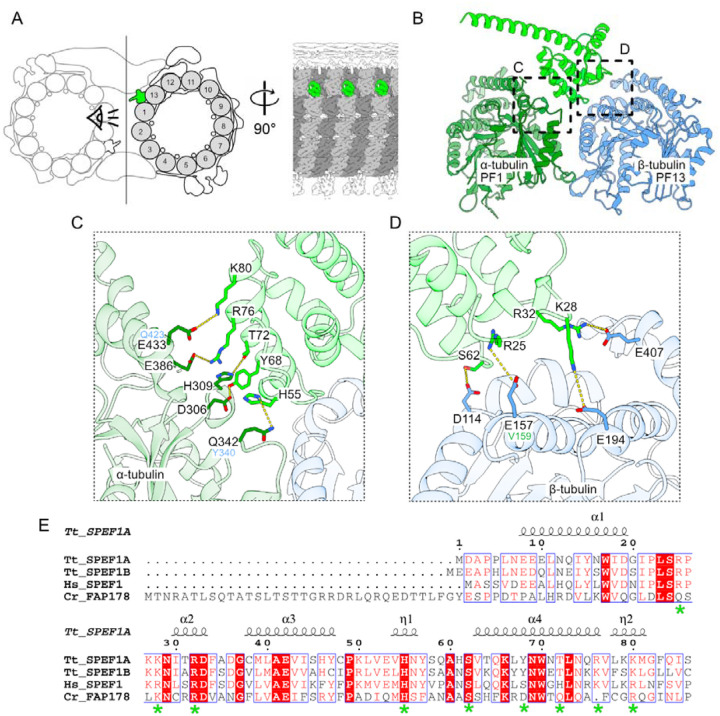
Figure 5: SPEF1 is a seam-binding protein.
(A) Diagram of the tip CP highlighting SPEF1A (green). α-Tubulin is light gray, and β-tubulin is dark gray. (B) Model of SPEF1A interacting with tubulin at the seam. (C, D) Amino acids that likely mediate the interaction between α-(C) and β-(D) tubulin. The equivalent amino acids in β- and α-tubulin are shown in blue in (C) and in green in (D), respectively. (E) Alignment of Tetrahymena SPEF1A1–86, SPEF1B, human SPEF1 (UniProtID Q9Y4P9) and Chlamydomonas FAP178 (UniProtID A0A2K3D8Z6). The amino acids shown in C and D are highlighted with a green asterisk.

