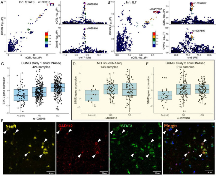
Fig. 6. Examples of COLOC results.
(A, B) The locus-compare scatter plot for the association signals at STAT3 and IL7 in the inhibitory neurons.
(C, D, E) Expression quantitative trait loci (eQTL) box plots of associations between genotype rs1026916 and STAT3 expression in inhibitory neurons using snucRNAseq data from Fujita et al. (CUMC study 1), Mathys et al. (MIT cohort), and our in-house multiome datasets (CUMC study 2).
(F) Immunohistochemistry of DLPFC in human MS brain tissue, stained for STAT3 (green), GAD1/2 (red), and NeuN (yellow), with DAPI (blue) to visualize nuclei. Expression of STAT3 was observed in NeuN+GAD1/2+ neurons. White triangles highlight the colocalization of DAPI, STAT3, GAD1/2, and NeuN. Scale bar, = 50 μm.

