. 2024 Dec 13;13:RP101269. doi: 10.7554/eLife.101269

Figure 3. Heavy chain-hyaluronic acid/pentraxin 3 (HC-HA/PTX3) inhibited heat hypersensitivity in wild-type (WT) mice after plantar-incision and attenuated dorsal root ganglion (DRG) neuron activation.

(A) Left: Intra-paw injection of HC-HA/PTX3 (10 μg or 20 μg, 20 μl), but not vehicle (saline), increased paw withdrawal latency (PWL) to heat stimulation in naive WT mice. N = 8–11/group. Right: Intra-paw injection of HC-HA/PTX3 (10 μg or 20 μg, 20 μl) dose-dependently attenuated the heat hypersensitivity during Days 2–4 after plantar-incision. N = 9–16/group. (B) Right: Intra-paw injection of HC-HA/PTX3 (20 μg, 20 μl) showed superior anti-hyperalgesic effect compared to high-molecular-weight hyaluronan (HMW-HA) (20 μg, 20 μl) alone and the mixture of HMW-HA (20 μg) and HC1 (720 ng) during Days 2–4 after plantar-incision. Left: Analyzing the area under the curve (AUC) to assess the anti-hyperalgesic effect of each group. N = 5–9/group. (C) HC-HA/PTX3 inhibited the calcium responses evoked by capsaicin (a TRPV1 agonist, 0.3 μM) in WT DRG neurons. HC-HA/PTX3 alone did not evoke [Ca2+]i elevation. Pretreatment (20 min) of HC-HA/PTX3 (15 μg/ml, bath application) reduced capsaicin-evoked [Ca2+]i rising. (D) The quantification of [Ca2+]i rising evoked by capsaicin in DRG neurons pretreated with the vehicle, HC-HA/PTX3 (15 μg/ml), or HMW-HA (15 μg/ml). N = 109–170 neurons/group. (E) Left: Traces show that the β-alanine (a MrgprD agonist, 1 mM) evoked an increase in [Ca2+]i, which was also inhibited by HC-HA/PTX3. Right: The quantification of evoke [Ca2+]i rising by β-alanine. N = 10–25 neurons/group. (F) Left: Traces show that cinnamaldehyde (a TRPA1 agonist, 1 mM) evoked an increase in [Ca2+]i, which was inhibited by HC-HA/PTX3. Right: The quantification of evoke [Ca2+]i rising by cinnamaldehyde. N = 15–35 neurons/group. (G) An example trace of membrane potential (Vm) which changed from resting level (−60 mV) toward a more hyperpolarized state after HC-HA/PTX3 (10 μg/ml) in a small DRG neuron (insert, scale bar: 25 μm). Vm returned to pre-drug level after washout. DRG neurons were categorized according to cell body diameter as <20 μm (small), 20–30 μm (medium), and >30 μm (large). (H) Example traces of action potentials (APs) evoked by injection of current in small DRG neurons 5 min after bath application of vehicle or HC-HA/PTX3 (5, 25 μg/ml). (I) HC-HA/PTX3 concentration-dependently altered the intrinsic membrane properties of small DRG neurons. Quantification of the resting membrane potential (RMP) before and at 5 min after bath application of vehicle or HC-HA/PTX3 (5, 10, and 25 μg/ml). N = 4–7/group. (J) Quantification of rheobase in small DRG neurons at 5 min after vehicle or HC-HA/PTX3. The rheobase after the drug was normalized to pre-drug value. N = 5–7/group. Data are mean ± SEM. (A, B: right) Two-way mixed model analysis of variance (ANOVA) followed by Bonferroni post hoc test. *p < 0.05, **p < 0.01, ***p < 0.001 versus vehicle; #p < 0.05, ##p < 0.01, ###p < 0.001 versus pre-drug. (B: left, C) One-way ANOVA followed by Bonferroni post hoc test. ***p < 0.001 versus vehicle; ##p < 0.01 versus other groups. (E, F) Paired t-test. ***p < 0.001 versus vehicle. (I, J) Two-way mixed model ANOVA followed by Bonferroni post hoc test. *p < 0.05, **p < 0.01 versus pre-drug.

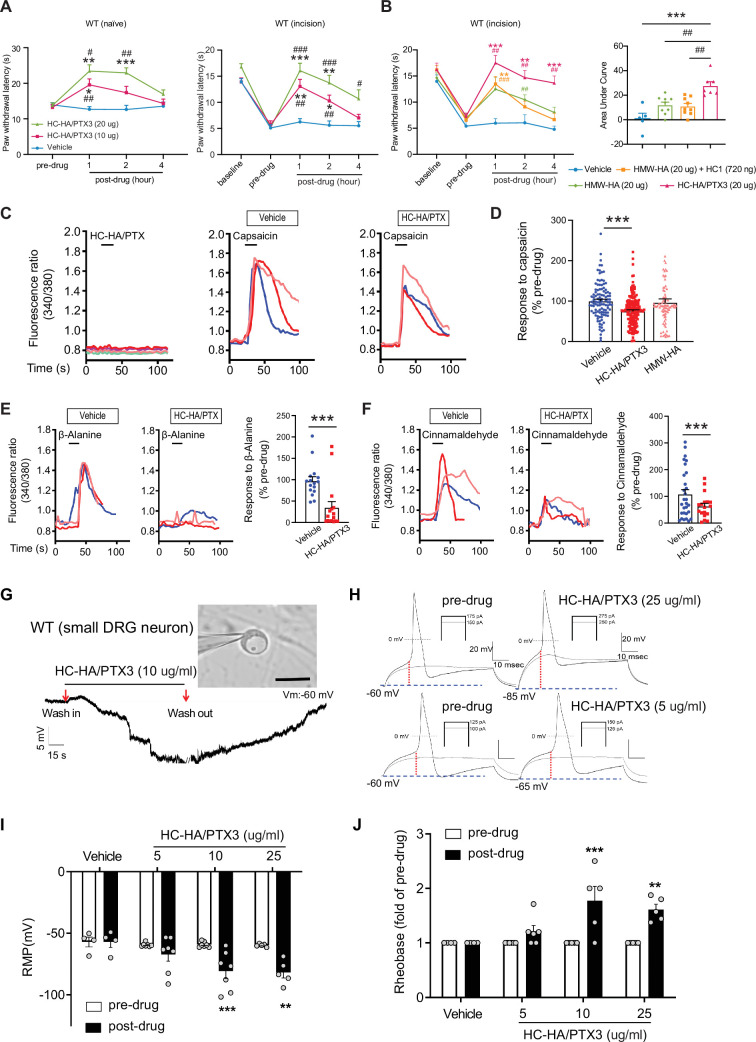
Figure 3—source data 1. Numerical source data files for Figure 3.

Figure 3.

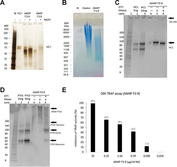
Figure 3—figure supplement 1. Purification and characterization of heavy chain-hyaluronic acid/pentraxin 3 (HC-HA/PTX3).

Figure 3—figure supplement 1.

(A) HC-HA/PTX3 was prepared from the human amniotic membrane by two runs (AM2P F3-9) or four runs (AM4P F3-9) of CsCl/4 M GnHCl ultracentrifugation and was then analyzed by silver staining. Each lane was loaded with 0.25 µg of HA without or with 100 mM NaOH treatment (25°C, 1 hr) to cleave the bond between HA and HC1. (B) HC-HA/PTX3 (AM4P F3-9) purified from the amniotic membrane was electrophoresed on 0.5% agarose gel and stained with All-stains dye. Healon as a high-molecular-weight (HMW) HA control and HC-HA/PTX3 were loaded at 10 µg HA/lane; M: HA molecular weight ladder. (C, D) HC1 and PTX3 in HC-HA/PTX3 were detected by western blot using respective antibodies without (−) or with (+) hyaluronidase (HAase) treatment to release HC1 or HMW-HA-PTX3, of which the latter can then be resolved into dimer or monomer without (−) or with (+) reduction with DTT. (E) Cloned murine RAW264.7 monocytes were seeded at 1 × 10⁴ cells/cm2 in MEM α/10% fetal bovine serum (FBS) and differentiated into multi-nucleated osteoclasts with 25 ng/ml RANKL as the positive control and treated with HC-HA/PTX3 at different concentrations (0.024–25 µg/ml) for 3 days. The inhibition of TRAP activity in cell lysates was calculated as a percentage (%) of that of the positive control (shown on the top of each bar).
Figure 3—figure supplement 1—source data 1. PDF file containing original western blots for Figure 3—figure supplement 1, indicating the relevant bands and treatments.
Figure 3—figure supplement 1—source data 2. Original files for western blot analysis displayed in Figure 3—figure supplement 1.
Figure 3—figure supplement 1—source data 3. Numerical source data files for Figure 3—figure supplement 1.
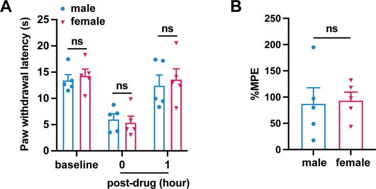
Figure 3—figure supplement 2. Heavy chain-hyaluronic acid/pentraxin 3 (HC-HA/PTX3)-induced comparable inhibition of heat hyperalgesia in male and female mice after plantar-incision.

Figure 3—figure supplement 2.

(A) Paw withdrawal latency at different time points of male and female mice is included in Figure 3B (N = 5/sex) (B) The percentage of maximal possible effects (%MPE) 1 hr post-drug were calculated for male and female mice included in Figure 3B. %MPE = 1 − (baseline − post-drug)/(baseline − pre-drug). Data are mean ± SEM. (A) Two-way mixed model analysis of variance (ANOVA) followed by Bonferroni post hoc test. *p < 0.05 versus male. (B) Unpaired t-test.
Figure 3—figure supplement 2—source data 1. Numerical source data files for Figure 3—figure supplement 2.
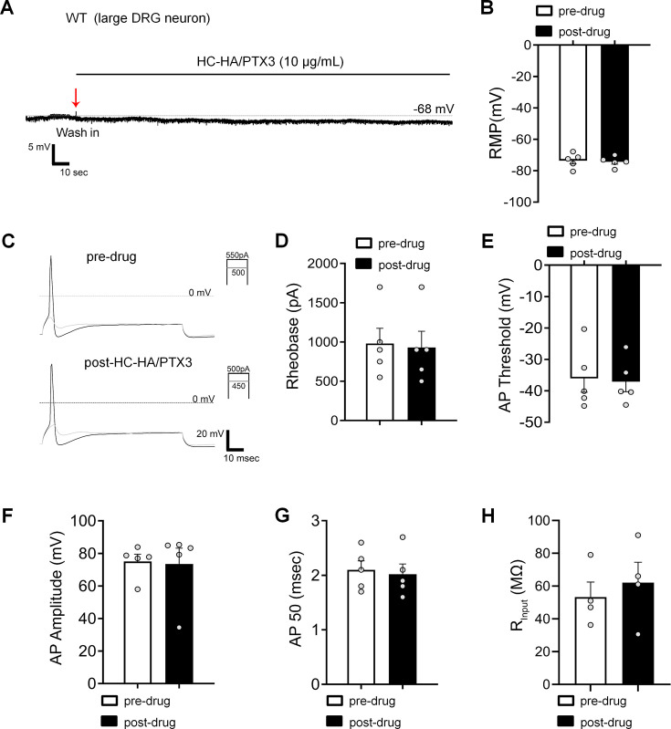
Figure 3—figure supplement 3. Heavy chain-hyaluronic acid/pentraxin 3 (HC-HA/PTX3) did not affect the excitability of large dorsal root ganglion (DRG) neurons in wild-type (WT) mice after the plantar-incision.

Figure 3—figure supplement 3.

(A) The representative trace of membrane potential was recorded under current-clamp conditions before and after HC-HA/PTX3 (15 µg/ml) treatment in a large DRG neuron of WT mice. Neurons were categorized according to cell body diameter as <20 μm (small), 20–30 μm (medium), and >30 μm (large). (B) The resting membrane potential (RMP) in large DRG neurons was not significantly changed at 5 min after HC-HA/PTX3 (10 μg/ml) treatment, compared to pre-drug (p = 0.41). N = 5. (C) Representative traces of rheobase measurements before and after HC-HA/PTX3 (10 μg/ml). Quantification of the rheobase (D, p = 0.09), action potential (AP) threshold (E, p = 0.78), AP amplitude (F, p = 0.8), AP duration (G, p = 0.41), and the mean input resistance (Rinput, H, p = 0.17) before and at 5 min after HC-HA/PTX3 (10 μg/ml) treatment. N = 5. Data are presented as mean ± SEM. (B, D–H) Paired t-test.
Figure 3—figure supplement 3—source data 1. Numerical source data files for Figure 3—figure supplement 3.