. 2024 Dec 13;13:RP101269. doi: 10.7554/eLife.101269

Figure 5. Heavy chain-hyaluronic acid/pentraxin 3 (HC-HA/PTX3) induced cytoskeletal rearrangement which contributed to its inhibition of INav and HVA-ICa.

(A) Example images show the distribution of F-actin and CD44 staining in small dorsal root ganglion (DRG) neurons of wild-type (WT) mice. Neurons were treated with bath application of vehicle (saline), high-molecular-weight hyaluronan (HMW-HA) (15 μg/ml), HC-HA/PTX3 (10, 15 μg/ml), or HC-HA/PTX3 (10, 15 μg/ml) combined with Latrunculin A (LAT-A, 1 μM) for 45 min. Scale bar: 5 μm. DRG neurons were categorized according to cell body diameter as <20 μm (small), 20–30 μm (medium), and >30 μm (large). (B) Quantification of sub-membranous F-actin polymerization and translocation of CD44 in small WT DRG neurons after drug treatment. N = 30–80/group. (C) Proliferation MTT assay showed a lack of neuronal toxicity from 0.5, 1, 2, 5, 10, and 15 μg/ml HC-HA/PTX3, compared to vehicle (100% viable cells). N = 6–12 repetitions/group. (D) Quantification of sub-membranous F-actin polymerization and translocation of CD44 in small DRG neurons. DRG neurons were electroporated with siRNA targeting Pfn1 (siPfn1) or non-targeting siRNA (siNT, control). Neurons were treated with vehicle (saline) or HC-HA/PTX3 (10 μg/ml) for 45 min. N = 70–111/group. (E) Changes in the sub-membrane distribution of F-actin and CD44 in WT DRG neurons treated with vehicle + control IgG (2 µg/ml), HC-HA/PTX3 (15 μg/ml) + control IgG (2 µg/ml), or HC-HA/PTX3 (15 μg/ml) + CD44 IgG (2 µg/ml) for 45 min. Scale bar: 5 μm. (F) Quantification of the sub-membrane F-actin and CD44 labeling in each group. (G) Infusion of LAT-A attenuated the inhibition of INav by HC-HA/PTX3 in WT DRG neurons. (a) Representative traces of INav after 5 min infusions of vehicle (top row) or LAT-A (bottom row, 0.5 nM) through the recording electrode, followed by bath application of HC-HA/PTX3 (10 µg/ml). Lumbar DRG neurons were harvested on Days 2–3 after plantar-incision. (b) There was a significant interaction between the variation produced by HC-HA/PTX3 (10 µg/ml) and test voltages (VTest) applied in vehicle-infused neurons, resulting in an overall INav inhibition (F(14,90) = 3.29, ***p < 0.001), and significantly decreased INav density (pA/pF) from VTest = −10 to +10 mV, as compared to pre-HC-HA/PTX3 treatment. N = 7/group. (c) HC-HA/PTX3 did not alter GNa/GNa max across the test voltages (F(9,60) = 0.44, p = 0.9) in vehicle-infused neurons. N = 7/group. (d) There was a significant interaction between the variation produced by HC-HA/PTX3 (10 µg/ml) and VTest applied in LAT-A-infused neurons, resulting in overall INav increase (F(14,120) = 1.87, *p < 0.05) and increased INav density (pA/pF) from VTest = −10 to 0 mV, as compared to pre-HC-HA/PTX3. N = 9/group. (e) HC-HA/PTX3 significantly increased the GNa/GNa max at VTest = −20 mV in LAT-A-infused neurons (*p < 0.05, N = 9/group). (H) LAT-A attenuated the inhibition of HVA-ICa by HC-HA/PTX3 in WT DRG neurons. (a) Representative traces of HVA-ICa in small WT DRG neurons after 5 min infusions of vehicle (top row) or LAT-A (bottom row, 0.5 nM), followed by bath application of HC-HA/PTX3 (10 µg/ml). (b) In vehicle-infused neurons, HC-HA/PTX3 (10 µg/ml) significantly decreased HVA-ICa (F(1,12) = 6.52, *p = 0.02) and HVA-ICa conductance (I/Imax) from VTest = −40 to +10 mV, as compared to pre-HC-HA/PTX3. N = 7. (c) HC-HA/PTX3 did not alter the channel open probability (Po) in vehicle-infused neurons (p = 0.82, N = 7). (d) In LAT-A-infused neurons, HC-HA/PTX3 only modestly reduced HVA-ICa conductance across test voltages applied (F(1,12) = 0.27, p = 0.6, N = 8). (e) HC-HA/PTX3 did not alter Po in LAT-A-infused neurons (p = 0.94, N = 8). Data are mean ± SEM. (B–D, F) One-way analysis of variance (ANOVA) followed by Bonferroni post hoc test. *p < 0.05, ***p < 0.001 versus vehicle; #p < 0.05, ###p < 0.001 versus indicated group. (G, H) Two-way repeated measures ANOVA with Holm–Sidak post-test. *p < 0.05, **p < 0.01, ***p < 0.001, ****p < 0.0001 versus vehicle infusion or LAT-A infusion group.

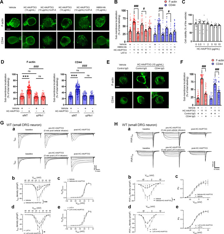
Figure 5—source data 1. Numerical source data files for Figure 5.

Figure 5.

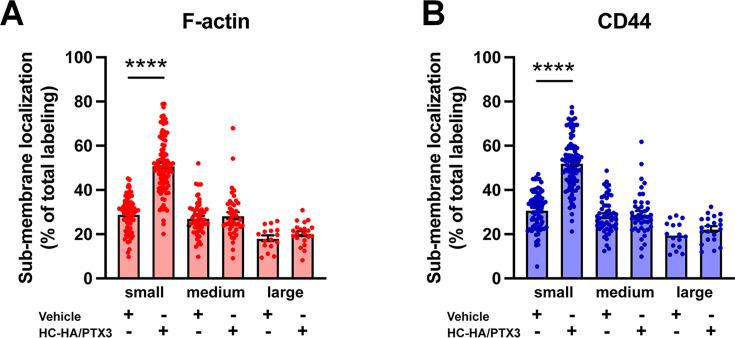
Figure 5—figure supplement 1. Differential effects of heavy chain-hyaluronic acid/pentraxin 3 (HC-HA/PTX3) on sub-membranous F-actin polymerization and translocation of CD44 in small, medium, and large wild-type (WT) dorsal root ganglion (DRG) neurons.

Figure 5—figure supplement 1.

(A) Quantification of sub-membranous F-actin polymerization and (B) translocation of CD44 in different sizes of WT DRG neurons after HC-HA/PTX3 (10 μg/ml) or the vehicle (saline) treatment. DRG neurons were categorized according to cell body diameter as <20 μm (small), 20–30 μm (medium), and >30 μm (large). HC-HA/PTX3 increased sub-membranous F-actin polymerization and translocation of CD44 exclusively in small neurons. N = 16–91/group. Data are mean ± SEM. One-way analysis of variance (ANOVA) followed by Bonferroni post hoc test. ****p < 0.0001 versus vehicle.
Figure 5—figure supplement 1—source data 1. Numerical source data files for Figure 5—figure supplement 1.
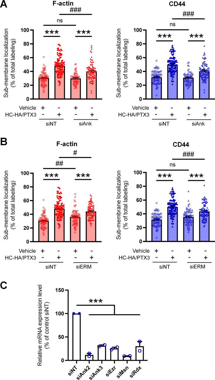
Figure 5—figure supplement 2. Quantification of sub-membranous F-actin polymerization and translocation of CD44 in small wild-type (WT) dorsal root ganglion (DRG) neurons in each group.

Figure 5—figure supplement 2.

(A) DRG neurons were electroporated with siRNAs specifically targeting Ank2 and Ank3 (siAnk), (B) and those targeting Ezr, Rdx, and Msn (siERM) complex. Neurons were treated with a bath application of vehicle (saline) or heavy chain-hyaluronic acid/pentraxin 3 (HC-HA/PTX3) (10 μg/ml) for 45 min. N = 59–114/group. (C) The mRNA expression of Ank2, Ank3, Ezr, Msn, and Rdx in DRG neurons electroporated with specific siRNAs were assayed by qPCR. N = 2. Data are mean ± SEM. One-way analysis of variance (ANOVA) followed by Bonferroni post hoc test. ***p < 0.001 versus vehicle; #p < 0.05, ##p < 0.01, ###p < 0.001 versus indicated group.
Figure 5—figure supplement 2—source data 1. Numerical source data files for Figure 5—figure supplement 2.
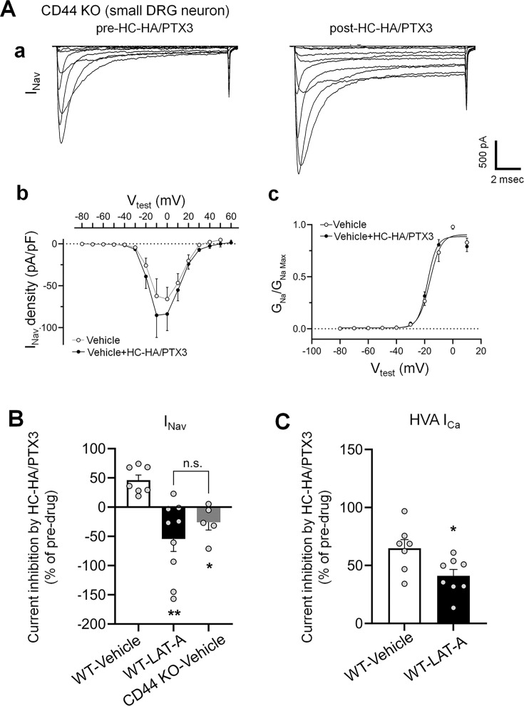
Figure 5—figure supplement 3. The inhibitions of INav by heavy chain-hyaluronic acid/pentraxin 3 (HC-HA/PTX3) in small dorsal root ganglion (DRG) neurons were diminished in CD44 KO mice and were inhibited by a pretreatment of LAT-A in neurons from WT mice.

Figure 5—figure supplement 3.

(A) HC-HA/PTX3 (10 µg/ml) did not inhibit INav in small DRG neurons from CD44 KO mice. (a) Representative traces of INav in a CD44 KO neuron before and at 5 min after bath application of HC-HA/PTX3 (10 µg/ml). (b) There was no significant change in INav density (pA/pF) before and after HC-HA/PTX3 (10 µg/ml) treatment in CD44 KO neurons. (c) HC-HA/PTX3 did not alter GNa/GNa max across the test voltages in CD44 KO neurons. N = 5. (B) Changes of INav after bath application of HC-HA/PTX3 (10 µg/ml) in vehicle-infused (N = 7) and LAT-A-infused small WT DRG neurons (p < 0.01, N = 9), and in vehicle-infused CD44 KO neurons (N = 5). DRG neurons were infused with the vehicle or LAT-A (0.5 nM) through the recording electrode, followed by bath application of HC-HA/PTX3 (10 µg/ml) 5 min later. The lumbar DRG neurons were harvested on Days 2–3 after the plantar-incision. DRG neurons were categorized according to cell body diameter as <20 μm (small), 20–30 μm (medium), and >30 μm (large). Data are mean ± SEM. One-way analysis of variance (ANOVA) with Holm–Sidak post-test. *p < 0.05, **p < 0.01 versus WT-vehicle pretreatment group. (C) The inhibition of HVA-ICa by HC-HA/PTX3 (10 µg/ml) in vehicle-infused (N = 7) and LAT-A-infused small WT DRG neurons (p = 0.02, N = 8). Data are mean ± SEM. Unpaired Student’s t-test.
Figure 5—figure supplement 3—source data 1. Numerical source data files for Figure 5—figure supplement 3.
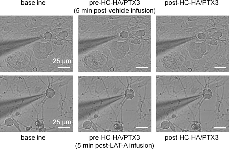
Figure 5—figure supplement 4. Intracellular infusion of LAT-A did not change the gross morphology of dorsal root ganglion (DRG) neurons in patch-clamp recordings.

Figure 5—figure supplement 4.

Example images show a small DRG neuron after infusion vehicle or Latrunculin A (LAT-A, 0.5 nM) through the recording electrode, followed by bath application of heavy chain-hyaluronic acid/pentraxin 3 (HC-HA/PTX3) (15 μg/ml). Scale bar: 25 µm. DRG neurons were categorized according to cell body diameter as <20 μm (small), 20–30 μm (medium), and >30 μm (large).