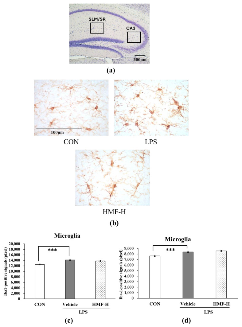
Figure 5.
The quantification of activated microglia between the stratum lacunosum moleculare (SLM) and stratum radiatum (SR) and in the CA3 region in the hippocampus of chronic inflammation model mice. (a) The locations of the captured images and quantification in the hippocampus are shown with squares. (b) Representative microglia pictures stained with an anti-Iba1 antibody in the SLM and SR. The scale bar represents 100 μm. (c) The quantification of Iba1-positive signals in the SLM and SR. (d) The quantification of Iba1-positive signals in the CA3 region. Data were analyzed by performing a one-way ANOVA followed by Dunnett’s multiple comparison test. *** p < 0.001 indicates a significant difference between CON and LPS. Values are presented as the means ± SEMs (n = 3–4 brain sections/mouse in each group).

