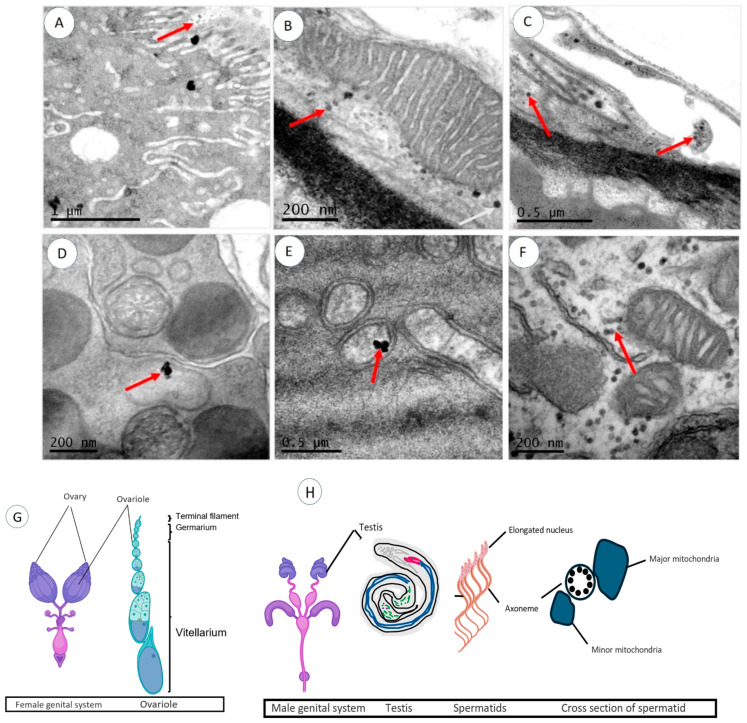
Figure 4.
TEM images of the reproductive organs of Drosophila. (A–C) Ultrathin section of Drosophila ovaries showing the distribution of AgNPs inside germanium (A), close to mitochondria (B), and in outer membranes (C). (D–F) Ultrathin sections of Drosophila testis showing the distribution of AgNPs inside spermatid cyst (D,E) and surrounding mitochondria (F). (G,H) Schematic drawings of Drosophila female and male genital systems, respectively. Red arrows point out AgNPs.

