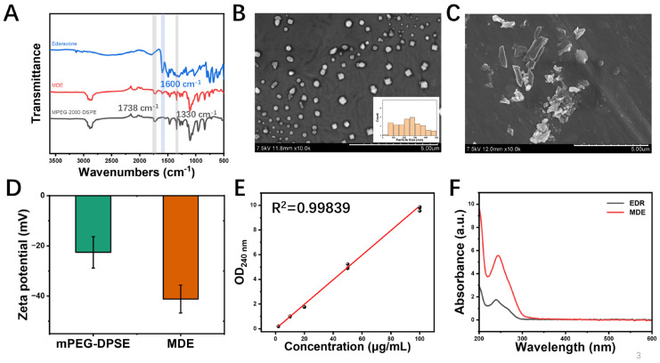
Figure 2.
Physical properties of MPEG-2000-DSPE-edaravone (MDE) micelles. (A) Fourier transform infrared spectroscopy of MDE, edaravone, and MPEG-2000-DSPE. (B) Scanning electron microscope image and the distribution of particle sizes of MDE; scale bar is 5 μm. (C) Scanning electron microscope image of edaravone (EDR); scale bar is 5 μm. (D) Zeta potential map of MDE and MPEG-2000-DSPE. (E) Standard curve of MDE. (F) Aqueous solubility curve of MDE (red) and edaravone (black).

