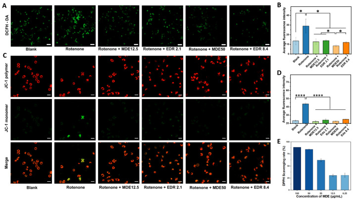
Figure 5.
Antioxidant effects of MPEG-2000-DSPE-edaravone (MDE) in Parkinson’s disease cell model. In the PD cell model, the logarithmic-growth-stage PC12 cells were treated with 1 μM rotenone for 24 h and further co-cultured with different concentrations of MDE or edaravone for another 48 h. Representative images were taken after incubating the vehicle medium, free edaravone, or MDE for 48 h. The intracellular reactive oxygen species (ROS) level was detected using a DCFH-DA fluorescent probe. (A) DCFH-DA staining. (B) Average fluorescence intensity of DCFH-DA. The mitochondrial membrane potentials were measured using the JC-1 assay kit. (C) JC-1 staining (mitochondrial membrane potentials). (D) Average fluorescence intensity of JC-1 monomer. (E) The antioxidant activity of MDE was evaluated using a DPPH radical scavenging assay. Data are presented as the mean ± SD of six independent experiments. * p < 0.05, **** p < 0.0001 versus control group. Scale bars in A are 50 μm. Scale bars in C are 20 μm.

