Figure 3.

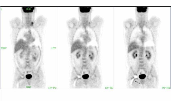
FDG-PET image of a patient with lymphoma involvement in the left cervical region. Note the periprosthetic artifact (arrow) in the region of the right hip in CT corrected images without segmentation.

FDG-PET image of a patient with lymphoma involvement in the left cervical region. Note the periprosthetic artifact (arrow) in the region of the right hip in CT corrected images without segmentation.