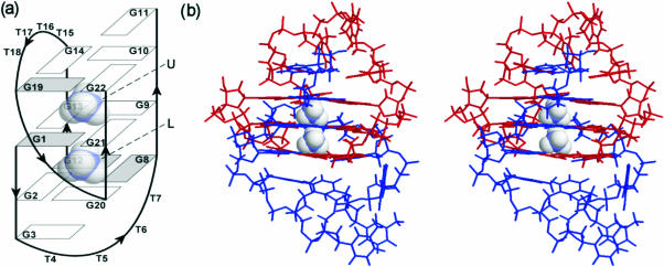
Figure 1.
(a) Schematic representation of topology of d(G3T4G4)2 G-quadruplex and its two cation binding sites labeled as U and L. The guanine bases are shown as rectangles, where filled rectangles represent syn nucleobases. (b) Stereo view of high resolution NMR structure (pdb = 1U64) of d(G3T4G4)2. Note a 180° rotation to the left with respect to (a). Strands G1–G11 and G12–G22 are colored blue and red, respectively.

