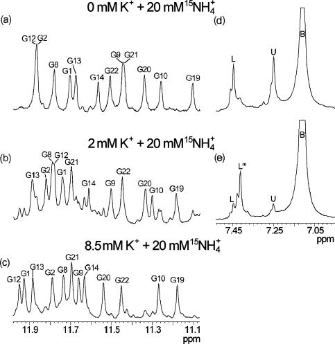
Figure 2.
Standard and 15N-filtered 1D 1H NMR spectra of d(G3T4G4)2 at 25°C and pH 5.0 in 10% 2H2O in the presence of 20 mM 15NH4Cl and 0 (a and d), 2 (b and e) and 8.5 mM (c) KCl. Assignments of individual imino resonances are indicated (a–c). Labels U (δ7.25 p.p.m.), L (δ7.45 p.p.m.), Lm (δ7.41 p.p.m.) and B (δ7.11 p.p.m.) indicate ions at distinct binding sites within the architecture of d(G3T4G4)2 and in bulk solution, respectively. Downfield shoulders of the main peaks in (d) and (e) are due to isotopomers like 15NH3D+. The oligonucleotide concentration was 3.5 mM in strand.

