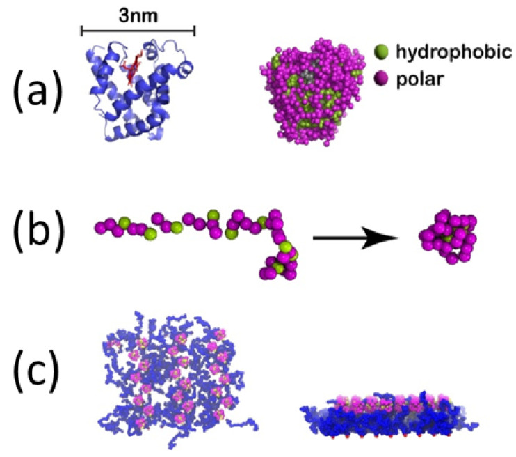
Figure 23.
(a) Myoglobin protein: (a-left image) Native structure showing the -helices. (a-right image) Coarse-grained model consisting of 151 amino acids, each one represented by a sphere. Polar amino acids are in red color, while hydrophobic (non-polar) ones are in green color. (b) Coarse-grained model of S25 protein consisting of 40 polar amino acids and 25 hydrophobic ones: (b-left image) Starting elongated configuration. (b-right image) Collapsed structure, where the hydrophobic sites have been brought inside the structure, leaving essentially all polar ones on its surface. (c) Planar polymer ‘mushroom’ in the case of 24 S25 proteins: (c-left image) Top view perpendicular to the surface. (c-right image) Side view (adapted from [277]).

