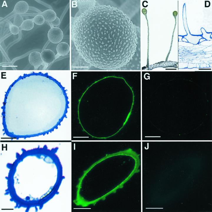
Figure 2.
Immunofluorescence localization of BAMT in snapdragon flower glands. A, Environmental scanning electron micrograph of glands within corolla tube of 7-d-old snapdragon flower. B, Environmental scanning electron micrograph showing the gland's head. C, Light microscopy photograph of glands within corolla tube. Fresh samples of snapdragon corolla tube were hand cut. D, Transverse section of glands within corolla tube of 7-d-old snapdragon flower. E, Cross-section through gland head within corolla tube of 7-d-old snapdragon flower. F, Cross-section through gland head within corolla tube of 7-d-old snapdragon flower treated with anti-BAMT antibodies and visualized by fluorescent FITC-conjugated secondary antibodies. G, Control section corresponding to F. Cross-section through gland head within corolla tube of 7-d-old snapdragon flower treated with preimmune serum and visualized by fluorescent FITC-conjugated secondary antibodies. H, Cross-section through gland stalk within corolla tube of 7-d-old snapdragon flower. I, Cross-section through gland stalk within corolla tube of 7-d-old snapdragon flower treated with anti-BAMT antibodies and visualized by fluorescent FITC-conjugated secondary antibodies. J, Control section corresponding to I. Cross-section through gland stalk within corolla tube of 7-d-old snapdragon flower treated with preimmune serum and visualized by fluorescent FITC-conjugated secondary antibodies. Scale bars = 100 μm (A and D), 20 μm (B, E–G, I, and J), 300 μm (C), and 10 μm (H).

