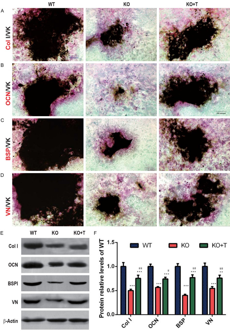
Figure 3.
Transplantation of wild-type BM-MSCs stimulated bone matrix protein synthesis and calcified nodule formation in BM-MSC cultures from 1α(OH)ase-/- recipients. (A) Representative micrographs of the resulting cells of bone marrow cell cultures derived from vehicle-treated wild-type (WT) and 1α(OH)ase-/- (KO) mice and BM-MSCs-transplanted 1α(OH)ase-/- mice (KO+T) with double staining using immunocytochemistry and Von Kossa for (A) type I collagen and von Kossa (Col I/VK), (B) osteocalcin and von Kossa (OCN/VK), (C) bone sialoprotein and von Kossa (BSP/VK), and (D) vitronectin and von Kossa (VN/VK). (E) Western blots of the cell extracts for expression of Col I, OCN, BSP and VN. β-actin was used as loading control. (F) Protein levels relative to β-actin protein were assessed by densitometric analysis and expressed relative to levels of WT mice. Each value is the mean ± SEM of determinations in 5 mice of each genotype. **: P<0.01; ***: P<0.001 relative to WT mice. #: P<0.05; ##: P<0.01, relative to KO mice.

