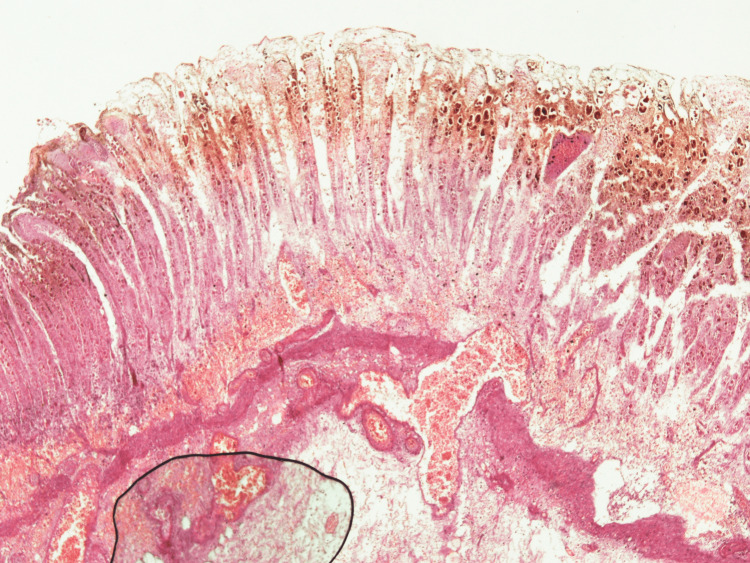
Figure 3. Histology of gastric tissue.
Full-thickness section of gastric tissue with transmural infarction, necrosis, and many hemosiderin-laden macrophages. Submucosal vessels were dilated and congested, and some were thrombosed. There was an extensive purulent process, extensively destroying the mucosa and submucosa.

