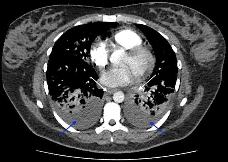
Figure 5. Chest CT scan.
Bilateral perihilar and centrilobular confluent consolidation along with small bilateral pleural effusions. Importantly, the pulmonary artery and its segmental branches were widely patent, showing no signs of pulmonary embolism or significant pericardial effusion. Additionally, there was no evidence of hilar or mediastinal lymphadenopathy, nor any notable bony abnormalities. These findings were consistent with bilateral bronchopneumonia and reactive effusion, effectively ruling out pulmonary embolism. (Arrows are added to highlight the bilateral perihilar consolidation located in the central part of both lungs near the hilum, annotated with white arrows. Additionally, the small bilateral pleural effusions are indicated at the lower posterior regions of both lungs, annotated with blue arrows.)

